
Mohs Surgery
What makes Mohs surgery unique is its meticulous nature, offering a high cure rate and minimizing damage to surrounding healthy tissue. This patient-friendly technique is particularly beneficial for treating skin cancers in sensitive areas where preserving cosmetic appearance is crucial.
The providers at SeaCoast Skin Surgery excel in Mohs surgery. Their expertise and focus on Mohs surgery have garnered international recognition, establishing them as leaders in advancing effective and patient-centered skin cancer treatments.
Examples of Mohs Surgery
What is Mohs Surgery?
This technique allows dermatologists, trained in Mohs surgery, to see beyond the visible disease, and to precisely identify and remove the entire tumor, leaving healthy tissue unharmed.
This procedure is most often used in treating three of the most common forms of skin cancer: melanoma, basal cell carcinoma and squamous cell carcinoma.
What are the Benefits of Mohs?
Mohs Surgery FAQs
By using detailed mapping techniques and complete microscopic control, the Mohs surgeon can pinpoint areas involved with cancer that are otherwise invisible to the naked eye. Therefore, even the smallest microscopic roots of cancer can be removed. The result is:
- The removal of as little normal skin as possible
- The highest possibility for curing the cancer
No. Mohs surgery is performed in a pleasant outpatient surgical suite and you may return home the same day. Hospital facilities are available if necessary.
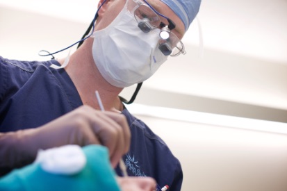
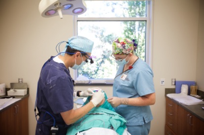
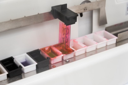
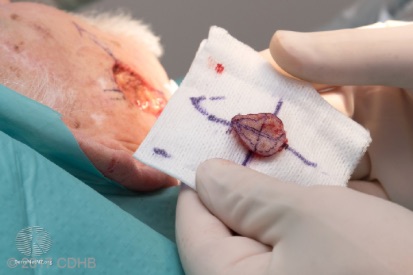
Your appointment will be scheduled early in the day. Our staff will escort you into a surgical suite where the surgeon will numb the area around the skin cancer. Once it is numb, the visible cancer and a thin layer of tissue will be removed. This tissue is carefully mapped and coded by the surgeon and taken to the adjacent laboratory where the technician will immediately process the microscope slides. You will have a temporary dressing placed over the wound and you will be free to return to the reception area.
The surgical procedure alone takes 10-15 minutes. However, it takes a minimum of 1 1/2 to 2 hours to prepare and microscopically examine the tissues of each layer. Several surgical stages and microscopic examinations may be required, and you will be asked to wait in the patient reception area between stages. Although there is no way to tell before surgery how many stages will be necessary, most cancers are removed in three stages or less.
We would like to make the time you spend with us as pleasant and comfortable as possible. You may want to bring reading material to occupy your time while waiting for the microscope slides to be processed and examined. You may want to bring a sweater, as the temperature in our office varies. Magazines and beverages will be available in the reception area. If your visit extends through the lunch hour, your companion may visit the hospital cafeteria and bring you a snack or lunch since you are asked not to leave the reception area of our office.
The most difficult part of the procedure is waiting for the results from the laboratory. Since we do not know in advance how much time is necessary to remove the cancer and repair the wound, we ask that you plan to be in the office the entire day and that you make no other commitments. Please be sure to inform your companion and/or driver of this.
Yes. Any form of treatment will leave a scar. However, because Mohs surgery removes as little normal tissue as possible, scarring is minimized. Immediately after the cancer is removed, we may choose (1) to leave the wound to heal itself, (2) to repair the wound with stitches, or (3) to reconstruct the wound with a skin graft or flap. This decision is based on the safest method that will provide the best cosmetic result.
After having skin cancer, you'll need Total Body Skin Exams (TBSEs) regularly, typically every 3 to 6 months. This frequent monitoring is crucial to catch any new or recurrent skin cancers early for effective treatment. Regular TBSEs help ensure your ongoing skin health and prevent potential complications. At Zitelli & Brodland, we'll work with you to ensure your custom skin care plan is in place for these exams.
The best protection from skin cancer is to avoid the harmful ultraviolet rays of the sun. Even if you tan easily, the sun can contribute to skin cancer in two ways. First, the sunlight damages the genes that control cell growth, and second, sunlight damages the body’s immune system so that early cancers grow unchecked by normal immune defense.
Minimize your exposure by:
- Using any sunscreen with a sun protective factor (SPF) of at least 30 and preferably with UVA/UVB protection when you spend any time in the sun.
- Avoid sun exposure during mid-day hours (10:00 am – 4:00 pm)
- Do not stay outdoors unprotected on cloudy days since the ultraviolet light penetrates easily through the clouds.
- If you follow this advice it may not be necessary to restrict your outdoor activities or change your lifestyle.
Mohs Surgery: The Most Effective Treatment for Skin Cancer
What to Expect at Your During Your Mohs Procedure
Once the obvious tumor is removed, Mohs surgeons:
- remove an additional, thin layer of tissue from the tumor site.
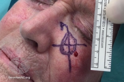
- create a “map” or drawing of the removed tissue to be used as a guide to the precise location of any remaining cancer cells.
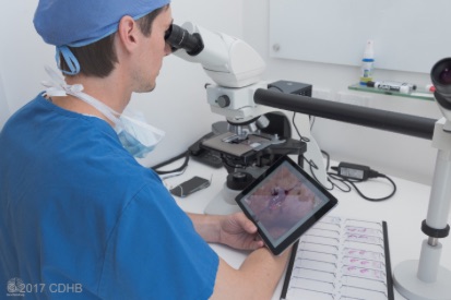
- microscopically examine the removed tissue thoroughly to check for evidence of remaining cancer cells.
- return to the specific area of the residual tumor indicated by the map
- remove another thin layer of tissue only from the specific area(s) where cancer cells were detected
- microscopically examine the newly removed tissue for additional cancer cells
How to Prepare for Mohs Surgery
These are general recommendations. We recommend working closely with your specific provider to plan and prepare for your Mohs surgery.
Planning for Recovery after Mohs Surgery
- If you live locally and feel comfortable coming alone to your appointment, have someone on standby to drive you home in case of an emergency.
- Plan to take it easy for a day or two, and possibly up to a couple of weeks if you have sutures, avoiding strenuous activities.
- And, follow your surgeon's post-operative care instructions diligently, which may include keeping the wound clean and dry.
After having one skin cancer, statistics show that you have a higher chance of developing a second skin cancer. You should have your skin checked by your referring dermatologist at least once a year for four years not only to examine the treated skin cancer but also to check for new skin cancers.
Featured Products for Sun Protection

EltaMD UV Physical SPF 41
For oil-free sun protection with just a touch of color, EltaMD's lightly tinted UV Physical is a healthy choice. This chemical-free mineral sunscreen has antioxidants to neutralize free radicals. Water-resistant UV Physical withstands water, humidity and perspiration. 3 oz

EltaMD UV Sheer Broad-Spectrum SPF 50
UV Sheer has a lightweight, hydrating formula that feels silky to the touch and light and airy on the skin. It goes on smooth absorbing quickly into skin and helps even out skin tone. This facial sunscreen offers up to 80 minutes of sweat and water-resistance making it the ideal choice for an active lifestyle, hot and humid weather and for anyone looking for a lightweight and hydrating sunscreen to live freely under the sun. UV Sheer is formulated to be compatible on all skin tones without leaving a white cast. 3.0 oz